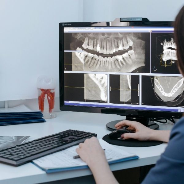
What your dentist can see during a visual exam is only the surface. Cavities between teeth, infections at the root tips, bone loss beneath the gums, and problems developing inside the jaw are invisible without imaging. Digital X-rays at Dental Sedation Ottawa use advanced sensor technology that produces detailed diagnostic images with up to 90% less radiation than traditional film — and results appear on screen instantly, so your dentist can diagnose and discuss findings during the same appointment. For patients who feel anxious about any dental procedure, including imaging, our sedation options make even routine X-rays comfortable.
Dental X-rays reveal what the naked eye simply cannot see. Up to 60% of the tooth structure is hidden beneath the gum line, and many of the most common dental problems develop in areas that are impossible to examine visually. Without imaging, early-stage cavities, infections, and bone loss can progress undetected until they cause pain, structural damage, or tooth loss.
X-rays allow us to detect cavities between teeth and beneath existing restorations long before they become visible or painful. They reveal infections at the root tips of teeth, where abscesses can develop silently for months. They show the extent of bone loss from periodontal disease — information that's critical for treatment planning. They identify impacted or developing wisdom teeth, cysts, tumours, and other pathology within the jaw.
For children and teens, X-rays monitor the development and eruption of permanent teeth, identify orthodontic concerns early, and detect decay in areas that are especially hard to clean. For adults, regular imaging catches problems at the stage when treatment is simplest, most conservative, and least expensive.
Skipping dental X-rays doesn't save money — it delays diagnosis. Problems caught early on an X-ray often need only a small filling or minor treatment. The same problem caught months or years later might require a crown, root canal, or extraction.
Sedation options ensure comfort for every patient — especially those with a sensitive gag reflex or dental anxiety.
We select the right type of imaging based on what we need to see — using the minimum radiation necessary for accurate diagnosis.
Digital X-ray technology represents a major advancement in both safety and diagnostic capability. Digital sensors require significantly less radiation to produce a clear image compared to traditional film — typically 70–90% less, depending on the type of image. This means patients receive a fraction of the radiation exposure that was standard just a generation ago.
To put dental radiation in perspective: a full set of digital dental X-rays (approximately 18 images) exposes you to roughly the same amount of radiation you receive from natural background sources during a single day of normal life. A single digital bitewing X-ray delivers less radiation than you'd receive during a short flight. Modern dental imaging is one of the lowest-radiation medical imaging procedures available.
Beyond reduced radiation, digital technology offers significant diagnostic advantages. Images appear on screen immediately — no waiting for film development. The images can be enhanced, magnified, and adjusted for contrast, making subtle problems easier to detect. Digital images can be easily shared with specialists for referrals and stored electronically for precise comparison over time.
We take radiation safety seriously and follow the ALARA principle — As Low As Reasonably Achievable. This means we use the minimum number of images needed for accurate diagnosis, optimize our equipment settings for the lowest effective radiation dose, use digital sensors that require far less radiation than film, and provide lead aprons and thyroid collars for all patients.
X-ray frequency is individualized based on your oral health status, risk factors, and dental history — not a one-size-fits-all schedule. Patients with stable oral health and low cavity risk may only need bitewing X-rays every 18–24 months. Higher-risk patients benefit from more frequent imaging to catch developing problems early.
For pregnant patients, we generally postpone routine X-rays until after delivery. However, if a dental emergency requires imaging for diagnosis and treatment, digital X-rays can be taken safely with appropriate shielding — the radiation dose is extremely low and the X-ray beam is directed away from the abdomen.
Many of the most serious dental conditions are invisible without imaging:
X-rays are covered as a diagnostic service by virtually all dental insurance plans. We verify your coverage before your appointment.
We provide direct billing to major insurers and accept the Canadian Dental Care Plan (CDCP) for eligible patients.
Coverage verified before your appointment — no surprises.
Sedation fees are additional to imaging costs: nitrous oxide $75–$150, oral sedation $150–$300.
Our patients consistently rate us 5 stars for gentle, anxiety-free care. Read verified patient experiences on Google.
View All Reviews on GoogleWhy patients choose Dental Sedation Ottawa for digital X-rays: